Introduction
The digestive system is made up of organs that digest food, assimilate its nutrients, and eliminate any leftover waste. The gastrointestinal (GI) tract, which connects the mouth to the anus, is essentially a long, continuous tube. For a variety of harmful bacteria, the alimentary canal serves as an immunological barrier. Gut-associated lymphoid tissue (GALT) and the various pH conditions that exist throughout the alimentary canal perform this role.
What is the Alimentary Canal?
Because of their complicated body plans, humans have a digestive tract with two openings: a mouth at one end and an anus at the other. The food material travels in a single path along the alimentary canal as it passes through several specialised organs. The Alimentary Canal is a similar tube whose main purpose is to facilitate food particle circulation and ultimately assist in nourishment.
Check out online study options are a great way to clear the science concepts you need. Study Science class 7th Lesson 2.
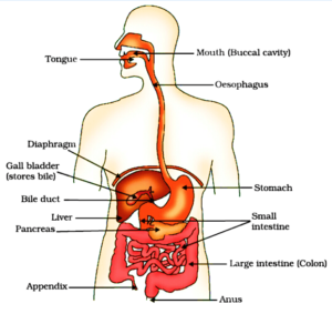
Parts of Alimentary Canal
Mouth: The mouth, also known as the oral cavity or buccal cavity, where the alimentary canal starts, contains teeth and a tongue that help break down food particles.
Pharynx: It is commonly referred to as the throat area. When food is swallowed, it travels via the pharynx.
Oesophagus: The throat and stomach are connected by a lengthy tube called oesophagus.
Stomach: The stomach is a structure that resembles an extended pouch that is situated between the oesophagus and the small intestine. Here, the food is transformed into a liquid suspension to facilitate absorption.
Small Intestine: As a result of the high rate of nutrient absorption in this area, it is sometimes referred to as the “workhouse” of digestion. It is the Alimentary Canal’s longest section.
Large Intestine: The alimentary canal’s terminus is located here. The “leftovers” in this area are used to absorb water and vital nutrients, which are eventually expelled through the anus
The Structure and Parts of Stomach
- The lower surface of the stomach’s curve to the left and the upper surface to the right is referred to as the lesser and larger curvatures, respectively.
- The fundus, body, and pylorus are its three parts. The fundus, which is the main part of the stomach, is elevated above the esophageal entrance.
- The stomach’s body and pylorus are it’s middle and base, respectively.
- The incisura angularis is the point where the body region and the proximal antrum converge.
- The muscularis mucosa, a thin layer of smooth muscle, is composed of the inner mucus epithelium, a bigger loose connective tissue called laminar propria, and the innermost layer of the gastrointestinal wall, known as the mucosa.
- Connective tissue, blood vessels, alveolar tissue, and Meiisner’s nerve plexus make up the submucosal layer.
Which are the Parts of the Alimentary Canal and What are their Functions?
| Parts of Alimentary Canal | Structure | Functions |
| Buccal Cavity |
|
|
| Pharynx |
|
|
| Oesophagus |
|
|
| Stomach |
|
|
| Small Intestine |
|
|
| Large Intestine |
|
|
Summary
Because of their complicated body plans, humans have a digestive tract with two openings: a mouth at one end and an anus at the other. The lower surface of the stomach’s curve to the left and the upper surface to the right are referred to as the lesser and larger curvatures, respectively. In the abdominal cavity, the small intestine looks like a lengthy, coiling loop. The lubricating mucus covered in faces is produced by the existing intestinal mucus glands.
Frequently Asked Questions (FAQs)
1. What is the Function of the Epiglottis?
Ans. Epiglottitis is a flap-like structure that covers the glottis, the windpipe’s entrance, when food is swallowed. This stops the food from choking and entering the windpipe.
2. What Function does Meiisner’s Nerve Plexus Serve?
Ans. Meissner’s nerve plexus controls the gastrointestinal tract’s secretions and local blood flow and aids in the start of the peristaltic movement.
3. Where are Delta Cells Found and what do they do?
Ans. The pancreatic islets of Langerhans contain delta cells. Somatostatin, a hormone that prevents the body from producing other hormones, is produced by these cells.
4. What Pigments are Present in Bile, and where do they Come From?
Ans. The bile pigments include greenish biliverdin and yellowish bilirubin. Dead red blood cells’ haemoglobin is degraded, and the bile pigments that result are expelled.
5. What Function does E. coli Serve in the Human Digestive System?
Ans. E. coli is a type of bacteria that helps with digestion, breaking down food particles for absorption in the small intestine and producing vitamin K.
 Mission Statement
“Empower every student to achieve full potential”
88Guru has been established with the social objective of making quality video-based learning material available to all Indian students. Technology, Connectivity and Social Media are rapidly changing the world of Education and we wish to lead the transformation of the tuition industry in India.
88Guru is the perfect complement to the current tuition model. 88Guru creates a wonderful opportunity for children and parents to bond while engaging in a valuable learning activity. It also provides the complete curriculum at your fingertips for those moments when you need some help at short notice. We believe that this mode of tuition could be transformational, adding hours to a child's day while providing complete control over the learning process.
Every course is taught by the best teachers from India's top schools and conducted in an engaging manner to keep students involved. The e-learning process consists of video-based instructions, computer-graded assignments, and a dashboard which allows the student and parent to track progress.