Introduction
The closed circulatory system of higher animals, such is made up of various blood vessels. The blood vessels make sure that every cell throughout the body receives an uninterrupted flow of oxygen-rich blood from the heart. The blood vessels have been specifically designed to carry one of the two types of blood-i.e oxygenated or deoxygenated blood to prevent their mixing. Deoxygenated blood is carried by the veins and venules, whereas oxygenated blood is carried by the arteries and arterioles. The third type of blood vessel, capillaries, are the smallest divisions and enable exchange between the blood and tissues. The blood vascular system is separated into two circuits: the systemic circuit, which provides healthy blood to the entire body, and sends impure blood to the heart, and the pulmonary circuit, which works between the heart and the lungs.
Arteries
- Blood is pumped from the heart through arteries and then into the body.
- The systemic arteries follow a path that starts in the left ventricle and travels through smaller distributions to every portion of the body.
- As a result, arteries play a crucial role in supplying the organs with blood and nutrients.
- Up to 10% of the blood in the body is stored in arteries at any given moment.
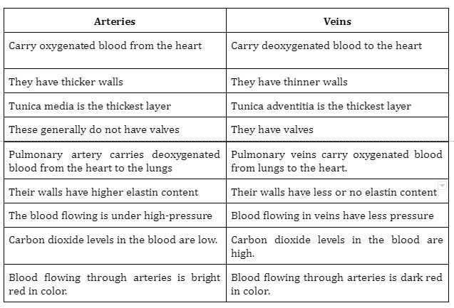
- Only one artery in the body, the pulmonary artery of the pulmonary circuit, transports deoxygenated blood from the heart’s right ventricle to the lungs.
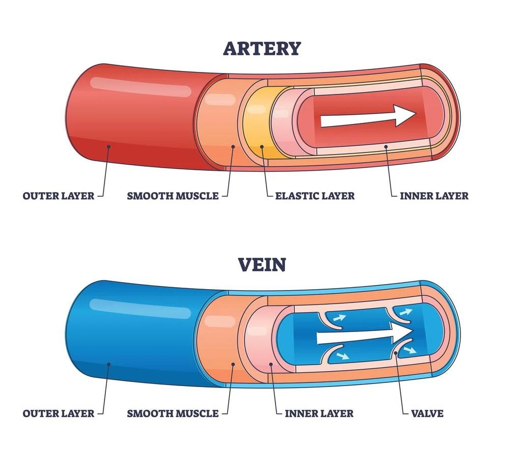
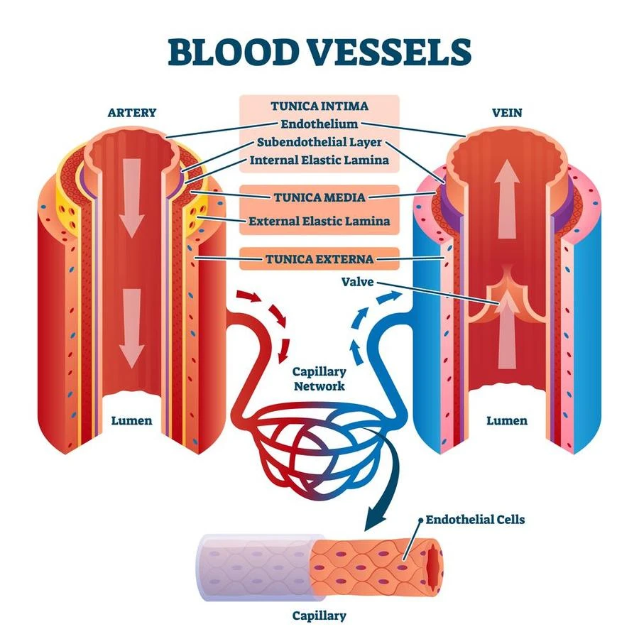
- The three layers of tissue that make up an arterial vessel—the tunica interna, tunica media, and the tunica externa/tunica adventitia—can vary in composition depending on the particular artery.
- The middle layer of the arterial wall, known as the tunica media, is the thickest layer and controls blood pressure by adjusting the vessel’s diameter.
- The type of vessel and the tasks it performs have a significant impact on the tunica media’s internal composition.
Types of Arteries and their Functions
There are three types of arteries. They are as follows-
- The Elastic Arteries
- They are the biggest and the closest to the heart; they are also referred to as conduit arteries. Examples include the aorta and the pulmonary arteries.
- The tunica media is strengthened by a lot of collagen and elastin to accommodate the blood surge since they get blood directly from the heart.
- As the heart beats, the artery walls can easily stretch because of elastin.
- Elastin also aids in keeping the pressure in the arteries constant.
- The elastic arteries buffer the cyclic changes that occur in the arterial blood pressure.
- The collagen fibers of the arterial wall bear the mechanical load at higher stresses, while the elastin fibers handle the low-pressure wall stretches.
- The Muscular Arteries
-
- The larger elastic arteries give rise to these medium-sized blood vessels.
More amount of smooth muscles and a reduced amount of elastin fibers are present in the tunica media of muscular arteries.
- These arteries are distributing arteries, which means they transport blood from the elastic arteries to the body’s organs and other regions.
- Actomyosin-mediated contractions and relaxations cause the smooth muscles in the tunica media to contract or relax as necessary. This helps in narrowing or expanding the arterial lumen to control blood pressure.
- Examples include the femoral artery, brachial artery, and radial artery.
- Arterioles
-
- These are the smallest of the arterial blood vessels, with an average internal diameter of only 30 𝝁m.
- Typically, there are only 1-2 layers of smooth muscles in the walls of these arterioles.
- Their primary duty is to transport blood to the capillaries from the major arteries.
- Due to its reduced diameter, which resists blood flow, arterioles serve as vascular resistors and control blood flow and blood pressure.
- They are also referred to as resistance vessels.
Veins
- The blood vessels known as veins are responsible for transporting tissues’ deoxygenated blood back to the heart for the purpose of reoxygenation.
- In the venous system, blood enters the venules from the capillaries, moves through the major veins, and eventually arrives at the heart. This is in contrast to blood flow occurring in the arteries.
- Up to 70% of the blood in the body is stored in the veins at any given time.
- The pulmonary veins bring oxygen-rich blood from the lungs to the heart, whereas the systemic veins transfer deoxygenated blood to the right atrium.
- The three layers of tissue that make up an veins are the tunica interna, tunica media, and the tunica externa/tunica adventitia.
- However, due to the decreased blood pressure, the diameter and number of smooth muscles in the veins are fewer.
- Due to the lower blood pressure that is flowing through them, the diameter and number of smooth muscles have decreased.
- Vein have valves that ensure blood flow in a forward direction only.
Types of Veins
- Systemic veins
-
- These veins transport blood from the bodily tissues to the right atrium since they are a part of the systemic circuit.
- The largest vein in the body, the vena cava, drains blood to the right atrium from both the lower and upper parts of the body.
- Pulmonary veins
-
- These veins are connected to the pulmonary circuit, which connects the heart and lungs.
- They deliver oxygenated blood from the lungs to the left atrium of the heart.
- Two pulmonary veins come from each lung, hence there are a total of 4 pulmonary veins.
- Superficial Veins
-
- These veins are numerous and located close to the skin’s surface.
- Examples: Saphenous veins, cephalic veins, basilic veins, etc.
- Deep Veins
- These veins are located deep inside the muscular tissues, and a systemic artery is present adjacent to them. For instance, the brachial vein lies next to the brachial artery, while the femoral vein lies next to the femoral artery.
- Venules
-
- These are the body’s tiniest veins, and they are responsible for transporting deoxygenated blood from capillaries to the major veins.
Difference between Arteries and Veins
Summary
- The cardiovascular system consists of an effective network of three different types of blood vessels, each with distinct purposes.
- The arteries transport blood rich in oxygen from the heart to the rest of the body.
- There are three different types of arteries: elastic arteries, muscular arteries, and arterioles.
- Veins can be deep-seated or superficial, and they return deoxygenated blood from the body to the heart.
- For every artery that supplies blood to an area, there is a corresponding vein that carries blood away.
Frequently Asked Questions
1. What color of blood is present in veins?
Ans: Blood present in the body is red in color. It is bright red when it has received oxygen i.e. while flowing through the arteries and dark red when it has lost oxygen i.e while flowing through the veins.
2. What are the 4 main arteries of the heart?
Ans: The four main arteries of the heart are-
- The right coronary artery
- The left main coronary
- The left anterior descending
- The left circumflex artery
3. What are the components of tunica adventitia?
Ans: The outermost tunica (layer) of a blood vessel, encircling the tunica media, is called the tunica externa , also referred to as the tunica adventitia. It is mainly made up of collagen.In arteries, it is supported by external elastic lamina.
 Mission Statement
“Empower every student to achieve full potential”
88Guru has been established with the social objective of making quality video-based learning material available to all Indian students. Technology, Connectivity and Social Media are rapidly changing the world of Education and we wish to lead the transformation of the tuition industry in India.
88Guru is the perfect complement to the current tuition model. 88Guru creates a wonderful opportunity for children and parents to bond while engaging in a valuable learning activity. It also provides the complete curriculum at your fingertips for those moments when you need some help at short notice. We believe that this mode of tuition could be transformational, adding hours to a child's day while providing complete control over the learning process.
Every course is taught by the best teachers from India's top schools and conducted in an engaging manner to keep students involved. The e-learning process consists of video-based instructions, computer-graded assignments, and a dashboard which allows the student and parent to track progress.